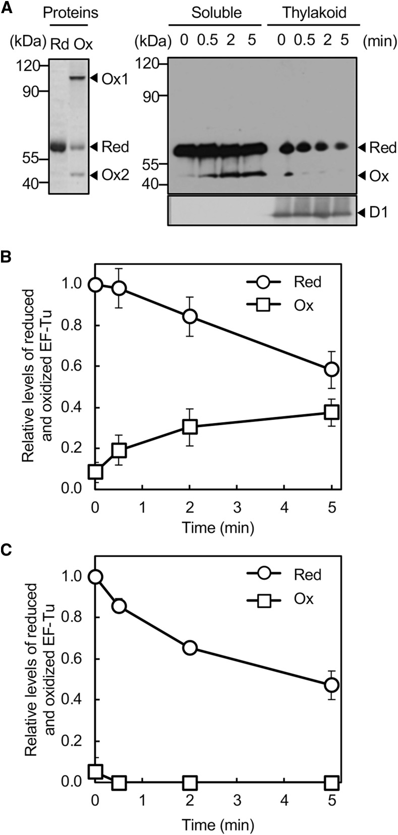
Figure 1.
Oxidation of EF-Tu under strong light in Synechocystis. A, Wild-type cells were exposed to strong light at 1000 μmol photons m−2 s−1 without aeration. Cells were harvested by filtration and lysed with glass beads. Cell extracts were separated into soluble and thylakoid membrane fractions and subjected to the thiol-modification assay with PEG-maleimide. Proteins were separated by nonreducing SDS-PAGE and EF-Tu was detected immunologically. For comparison, His-tagged EF-Tu protein that had been treated with 5 mm DTT (Red) or 1 mm H2O2 (Ox) was subjected to the thiol-modification assay and then to nonreducing SDS-PAGE. The D1 protein in membrane fractions was also detected immunologically. B, Quantitation of relative levels of reduced and oxidized EF-Tu in soluble fractions. C, Quantitation of relative levels of reduced and oxidized EF-Tu in thylakoid membrane fractions. Levels of reduced EF-Tu at 0 time in each fraction were taken as 1.0. Values are means ± sd (bars) of results from three independent experiments. The absence of bars in all figures indicates that the sd falls within the symbol. Ox1, oxidized dimer; Ox2, oxidized monomer; Red, reduced form.

