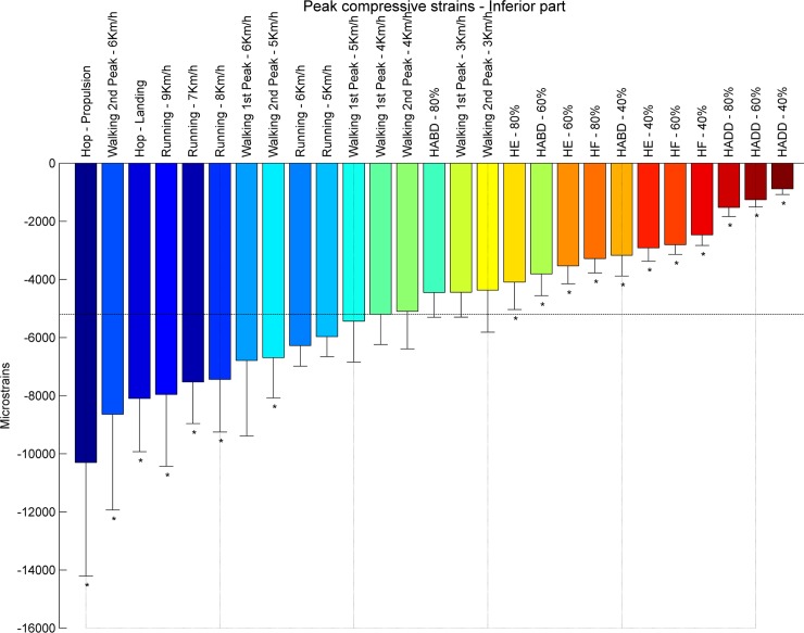
Fig 8. Ranking compression strains inferior part.
Average peak compressive strains in μstrains (εμ) in the inferior part of the femoral neck ranked from left to right for the highest (blue) to the lowest strain (red). Asterisks denote the exercises with significantly different peak compressive strains compared to walking at 4 km/h (1st peak) indicated by the horizontal line.

