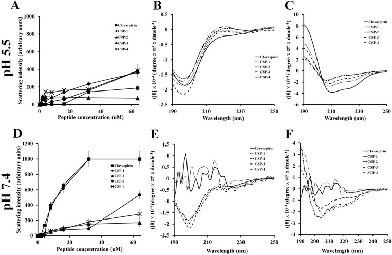
Figure 5. Aggregation of clavaspirin and its analogue peptides at pH 5.5 and pH 7.4.
Aggregation of clavaspirin and its analogue peptides. (A, B) Peptides incubated for 12 h at room temperature in 10 mM sodium phosphate buffer at pH 5.5 or pH 7.4 were monitored for structural changes using light scattering on an LS55 luminescence spectrometer (excitation 400 nm and emission 400 nm). Circular dichroism (CD) spectra analyses of peptides were incubated in 10 mM sodium phosphate (D, E left panel) or 30 mM SDS (C, F right panel). The secondary structures of CSP and its analogue peptides were determined using Far-UV CD spectra. All values represent the mean ± SD of three individual experiments (p < 0.05, one-way ANOVA).

