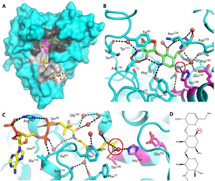
Fig. 1. Binary complexes of AAC-VIa.

(A) Surface representation of AAC-VIa–sisomicin (green carbon atoms) binary complex superimposed on the enzyme–acetyl-CoA (yellow carbon atoms) binary complex. The AAC-VIa surface cavities that bind sisomicin and coenzyme A (CoASH) are colored black and gray, respectively, whereas the catalytic triad is shown in magenta. (B) Close-up view of sisomicin (green carbon atoms) interaction with AAC-VIa. The site of antibiotic acetylation is circled in red. (C) Close-up view of acetyl-CoA (yellow carbon atoms) interaction with AAC-VIa. The acetyl donor is circled in red. Hydrogen bonds are represented as black dashed lines, and water molecules are shown as red spheres. The active-site residues have carbon atoms colored magenta. (D) Sisomicin with the site of acetylation (the N3 position of the unprimed ring) circled.
