Figure 3.

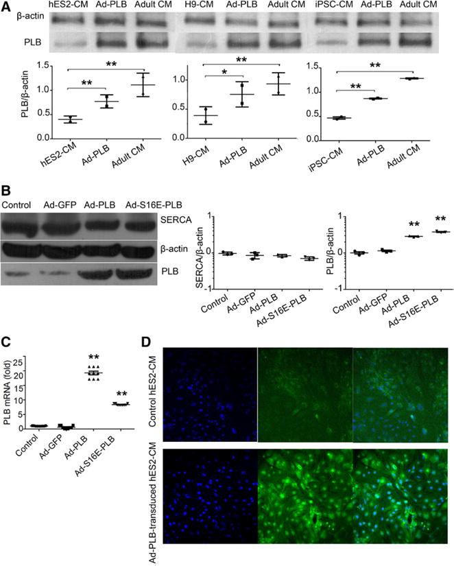
Effects of adenovirus (Ad)-green fluorescence protein (GFP), Ad-phospholamban (PLB), and Ad-S16E-PLB transduction on the expression levels of PLB, sarco/endoplasmic reticulum Ca2+-ATPase (SERCA). A, Western blot showing Ad-PLB transduction increased PLB protein expression in hES2-ventricular cardiomyocytes (vCMs; left), H9-vCMs (middle), and induced pluripotent stem cell (iPSC)-vCMs (right) but did not reach the adult level (n=2; P=0.226, 0.644, and 0.001 for hES2-vCMs, H9-vCMs, and iPSC-vCMs). B, Western blot analysis shows that the PLB protein levels of the Ad-PLB and Ad-S16E-PLB groups were significantly increased, but those for SERCA were not different from the untransduced and Ad-GFP–transduced groups (n=3). C, PLB transcript levels as gauged by quantitative polymerase chain reaction (n=3). D, Immunostaing images show that Ad-PLB–transduced hES2-vCMs were 100% positive: cell nucleus indicated by DAPI (left), PLB staining (middle), and merged image (right).
