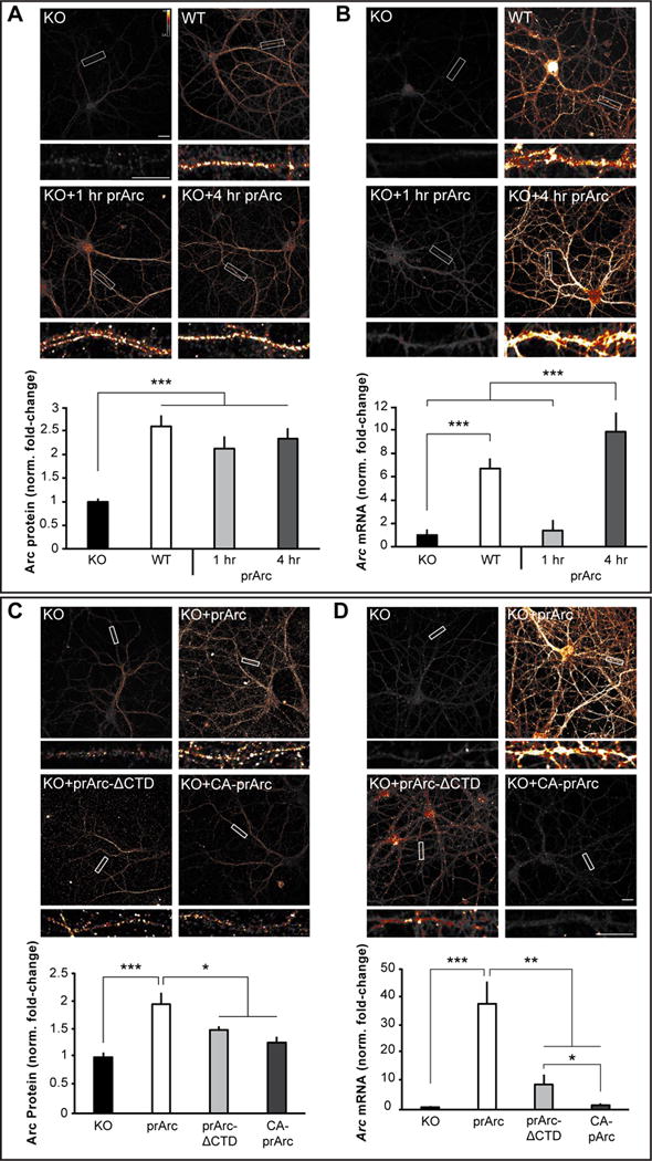
Figure 5. Arc capsids transfer Arc mRNA into neurons.

(A) Representative images of Arc ICC from DIV15 cultured hippocampal Arc KO neurons treated for 1 or 4h with 4μg prArc or WT control neurons. prArc-treated neurons showed increased dendritic Arc levels in untreated KO neurons. (B) Neurons were treated as in (A); representative images of Arc mRNA (FISH) are shown. 4h of prArc treatment significantly increased dendritic Arc mRNA levels. (C) Representative images of Arc ICC from DIV15 cultured hippocampal KO neurons treated with 4μg prArc, prArc-ΔCTD, or CA-prArc for 4h. KO neurons treated with prArc-ΔCTD and CA-prArc showed lower levels of Arc protein than prArc. (D) Neurons were treated as in (C); representative images of Arc mRNA are shown. Neurons treated with prArc-ΔCTD and CA-prArc showed lower levels of Arc mRNA than prArc. Dendritic segments boxed in white are shown magnified beneath each corresponding image. 30-μm segments of two dendrites/neuron were analyzed for integrated density measurements in all groups (n=10 neurons). Arc mRNA and Arc protein levels were normalized to untreated KO neurons and displayed as fold-change±SEM. Student’s t-test: *p<0.05. **p<0.01. ***p<0.001. Scale bars=10μm. Images are false-colored with the Smart LUT from ImageJ. All data are representative of 3–7 independent experiments using different protein preparations and cultures.
