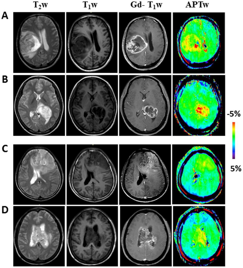
Fig. 3.
A, B Conventional and APTw MR images of two typical GBMs with an unmethylated MGMT promoter, illustrating the heterogeneous ring-enhancement characteristic of the disease on Gd-T1w image. APTw image demonstrated the lesion with ring-like, strong hyperintensity. C, D Conventional and APTw MR images of two typical MGMT promoter-methylated GBMs. Gd-T1w image demonstrated a patchy Gd-enhancing tumor mass. APTw image showed the masses as a hyperintense nodule (C) or scattered patch (D).

