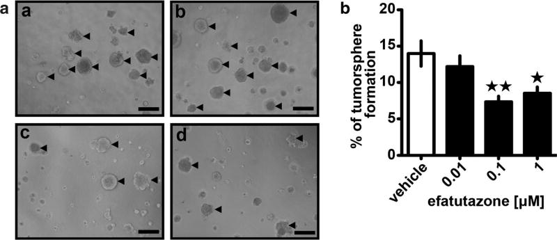
Fig. 3.
Efatutazone treatment leads to a reduction of MCFDCIS tumorsphere formation. a Representative phase-contrast images of MCFDCIS cultivated on Matrigel for 8 days in the presence of vehicle (a), 0.01 µM (b), 0.1 µM (c), and 1 µM efatutazone (d). Tumorspheres with diameter ≥ 100 µm are indicated by arrows. Scale bar = 200 µm. b Quantification of the percentage of MCFDCIS tumorspheres with diameter ≥ 100 µm treated with increased concentrations of efatutazone using Fiji software. Mean ± SEM (Student’s t test; n = 5; *p < 0.05 vs vehicle; **p < 0.01 vs vehicle)

