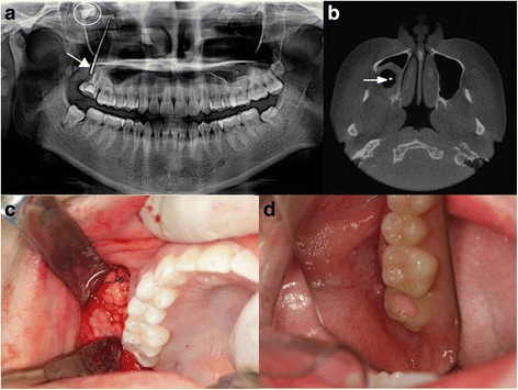
Fig. 1.

Case 1. a Panoramic view shows an oroantral fistula between the oral cavity and right maxillary sinus cavity (arrow indicates the gutta-percha cone). Additionally, full impaction of no. 18 was observed in the sinus cavity (circle). b Computed tomography showing right sinusitis and gutta-percha cone for examination (arrow). c The defect was reconstructed with pedicled buccal fat pad. d After 3 months, the operation site was successfully closed and well healed
