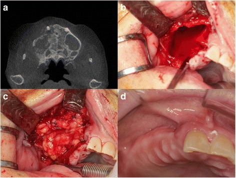
Fig. 2.

Case 2. a CT showed destruction of the right maxillary alveolar bone and right maxillary sinusitis. b Intraoperative view showed large oroantral defect after removal of inflammation tissue and sequestra. c The defect was covered with pedicled buccal fat pad with holding suture. d After 3 months, the operation site was successfully closed and well healed
