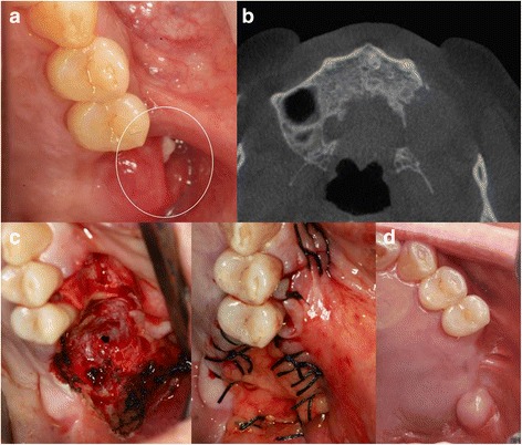
Fig. 3.

Case 3. a Preoperative oral examination showed a fistula on the gingiva due to recurrent ameloblastoma lesion (circle). b Preoperative CT showing destruction of alveolar bone on left maxillary posterior area. c During the surgery, a large defect was observed in the left maxillary posterior area due to the ameloblastoma. The pedicled buccal fat pad was used for coverage of the defect. The pedicled flap was exposed. d After 6 months, the site was well epithelized by soft tissue
