Figure 2.
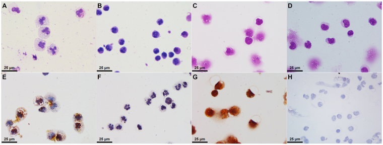
C-AgNP20 induced different cytomorphological alterations and intracellular distributions in cPMNs and cPBMCs. Representative cytological slides stained with Liu’s stain of cPMNs after 4 h of culture (A) with 50 μg/ml C-AgNP20 and (B) without C-AgNP20 (control), cPBMCs after 4 h of culture (C) with 50 μg/ml C-AgNP20 and (D) without C-AgNP20 (control); representative cytological slides stained with silver enhancement method of cPMNs after 4 h of culture (E) with 50 μg/ml C-AgNP20 and (F) without C-AgNP20 (control); cPBMCs after 4 h of culture (G) with 50 μg/ml C-AgNP20 and (H) without C-AgNP20 (control).

