Figure 1.
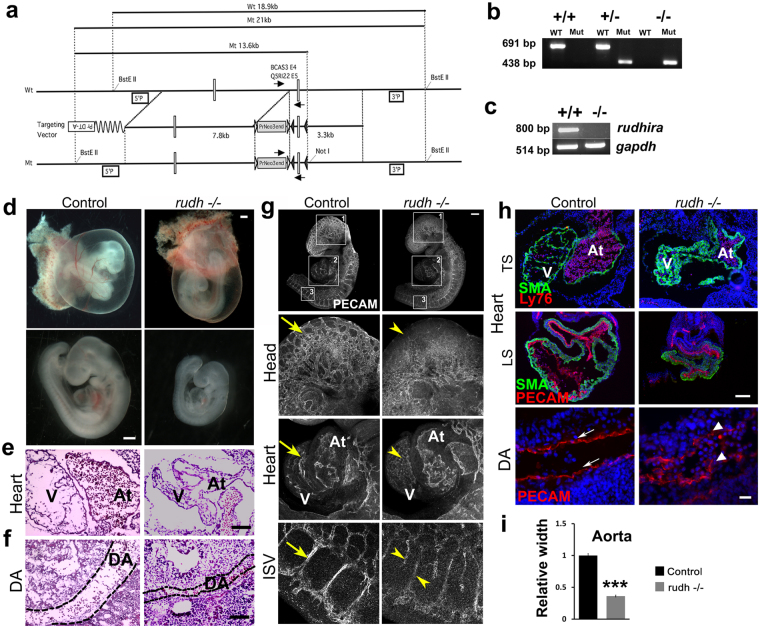
Rudhira is essential for development and cardio-vascular patterning. (a) Schematic showing strategy for generation of floxed allele of rudhira at exon 6. Rectangles: exons; black and white triangles: loxP and frt sequences respectively; 5’P and 3’P: probes for Southern blot analyses; arrows: genotyping primers. (b) PCR analysis showing genotype of control (+/+), heterozygous knock-out (+/−) and homozygous knock-out (−/−) embryos. (c) RT-PCR analysis showing rudhira mRNA expression in control (+/+) and homozygous knock-out rudh−/− (rudhfl/fl;CMVCre+) (−/−) embryos. GAPDH: loading control. (d–i) Analysis of control and knockout embryos at E9.5. (d) Unstained embryos, (e,f) Histological analysis comparing heart and dorsal aorta (DA). (g) Whole mount PECAM staining in control and rudh−/− embryos as indicated, with magnified view of head, heart and intersomitic vessels. (h) Immunostaining analysis of heart and dorsal aorta (DA) using myocardial marker SMA, primitive erythroid marker Ly76 and vascular markers PECAM. Arrows: normal vascular patterning; arrowheads: irregular and discontinuous vasculature. N = 5 embryos per genotype. (i) Graph shows quantitation of dorsal aorta width in the thoracic region. TS: Transverse section, LS: Lateral section, V: Ventricle, At: Atrium. Black dotted lines mark the boundary of DA. Scale bar: (d) 500 μm; (e,f) 100 μm; (g) 500 µm; (h) E9.5 heart: 100 μm, DA: 20 μm. The observed chi-square values for two degrees of freedom were 9.18 (E8.5), 13.08 (E9.5), 23.42 (E10.5), 42.96 (E11.5) and 37.5 (Postnatal).

