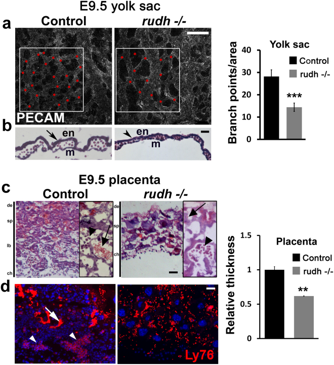
Figure 2.
Rudhira is required for extra-embryonic vascular patterning. (a) Morphology of whole mount PECAM/CD31 stained yolk sac Red dots: branch points. Graph shows quantitation of branch points (N = 5 yolk sacs). (b) hematoxylin-eosin stained yolk sac sections. en: endoderm, m: mesoderm. (c,d) Placental morphology seen by histological analysis (c) or immunofluorescence staining (d) in sections of control and rudh−/− as indicated. Boxed areas (in c) show magnified images of respective placenta. Immunostaining shows fetal and maternal blood cell pockets marked by Ly76. Nuclei are marked by DAPI (Blue). Arrows: maternal blood cells; arrowheads: fetal blood cells. de: decidua, sp: spongiotrophoblast, lb: labyrinth, ch: chorion. Graph shows quantitation of placental thickness. Results shown are a representative of at least three independent experiments with at least three biological replicates each. Statistical analysis was carried out using one-way ANOVA. Scale bar: (a,b) 200 µm; (c,d) 100 µm. *p < 0.05, **p < 0.01, ***p < 0.001.

