Figure 6.
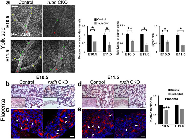
Endothelial deletion of rudhira leads to unpatterned extraembryonic vasculature. (a) Yolk sac vasculature marked by PECAM staining in control and rudhCKO (rudhfl/fl;TekCre+) at E10.5 and E11.5. Red dot: primary blood vessel; green dot: secondary blood vessel; yellow dot: tertiary blood vessel. Graphs show quantitation of number of secondary vessels, branch points and lumen size in control and rudhCKO at E10.5 and E11.5. (b–e) Placental morphology seen by histological analysis (b, d) or immunofluorescence staining (c,e) in sections of control and rudhCKO as indicated. Boxed areas show lower magnification of placentas (b,d). Immunostaining shows fetal and maternal blood cell pockets marked by Ly76. Nuclei are marked by DAPI (Blue). Arrows: maternal blood cells; arrowheads: fetal blood cells. de: decidua, sp: spongiotrophoblast, lb: labyrinth, ch: chorion. Graph shows quantitation of placental thickness. Results shown are a representative of at least three independent experiments with at least three biological replicates. Statistical analysis was carried out using one-way ANOVA. Scale bar: (a) 200 μm; (b–e) 100 µm. *p < 0.05, **p < 0.01, ***p < 0.001.

