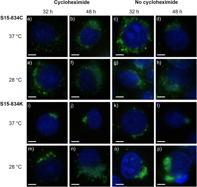
Figure 2.
Immunofluorescence images of S15-834C and S15-834K at 32 and 48 hours post infection (hpi). Shown are immunofluorescence images illustrating the morphology of S15-834C (a–h) and S15-834K (i–p) at 32 hpi (columns 1 and 3) and 48 hpi (columns 2 and 4) in the presence (columns 1–2) or absence (columns 3–4) of cycloheximide. Strains were grown at 37 °C (top line per strain; lines 1, 3,) and 28 °C (bottom lane per strain; lines 2, 4,). The size bar indicates 5 µm. Chlamydial inclusions are shown in green, the LLC-MK2 nuclei (DAPI) are shown in blue.

