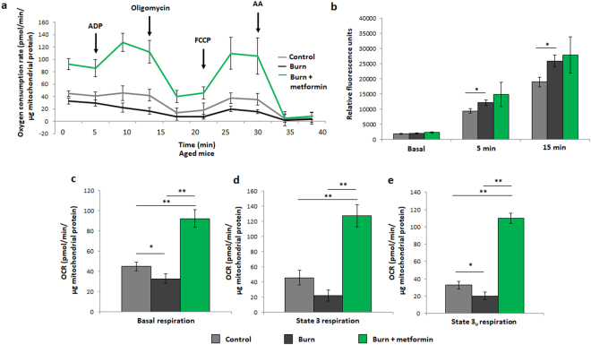
Figure 5.
Seahorse XF96 respirometry analysis of aged mice at day 7 post injury. (a) Respiration profiles of liver mitochondria from control mice (grey), burned mice (black) and burned mice with metformin treatment (100 mg/kg; green). (b) Production of reactive oxygen species as measured by oxidation of DCFDA in isolated mitochondria given substrate (5 mM pyruvate, 3 mM malate, 4 mM ADP); λ = 495 nm, λ’ = 529 nm. Basal (c), state 3 (d) and state 3 u (e) respiration parameters in isolated mitochondria as measured via Seahorse XF96 extracellular flux assays.  : control (n = 5); ■: burn (n = 5);
  : burn + metformin treatment (n = 6). Values are presented as mean ± standard deviation. *p ≤ 0.05; **p ≤ 0.01.

