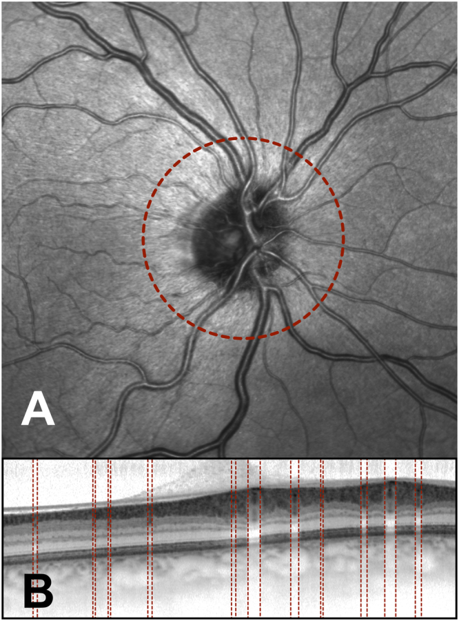
Figure 3.

Peripapillary spectral domain optical coherence tomography (SD-OCT) scan (B) and the linked confocal scanning laser ophthalmoscope (cSLO) fundus image (A) obtained from a subject who had not experienced a myocardial infarction at a young age (<50 years). The peripapillary OCT scanning circle was 3.5 mm in diameter and is shown in the cSLO image (red circle, A). The manually identified vessel borders are shown on the SD-OCT scan (red lines, B).
