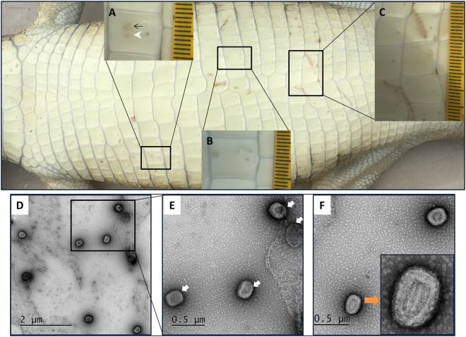
Figure 1.
Macroscopic (top panel) and transmission electron microscopic (bottom panel) analysis of saltwater crocodile tissues infected with poxvirus. Belly skin of juvenile saltwater crocodile showing poxvirus lesions as defined by Moore et al.3 (A) has an early active (black arrow) and active (white arrowhead) poxvirus lesion in the mid-scale region, (B) has an active lesion on the upper scale margin, and (C) shows poxvirus lesions along two linear blemishes ranging from the active to expulsion stages. Different stages of virus maturation (D–F) including immature virion (IV) (E, white arrow), and intracellular mature virion (IMV) (F, orange arrow) were imaged by transmission electron microscopy.

