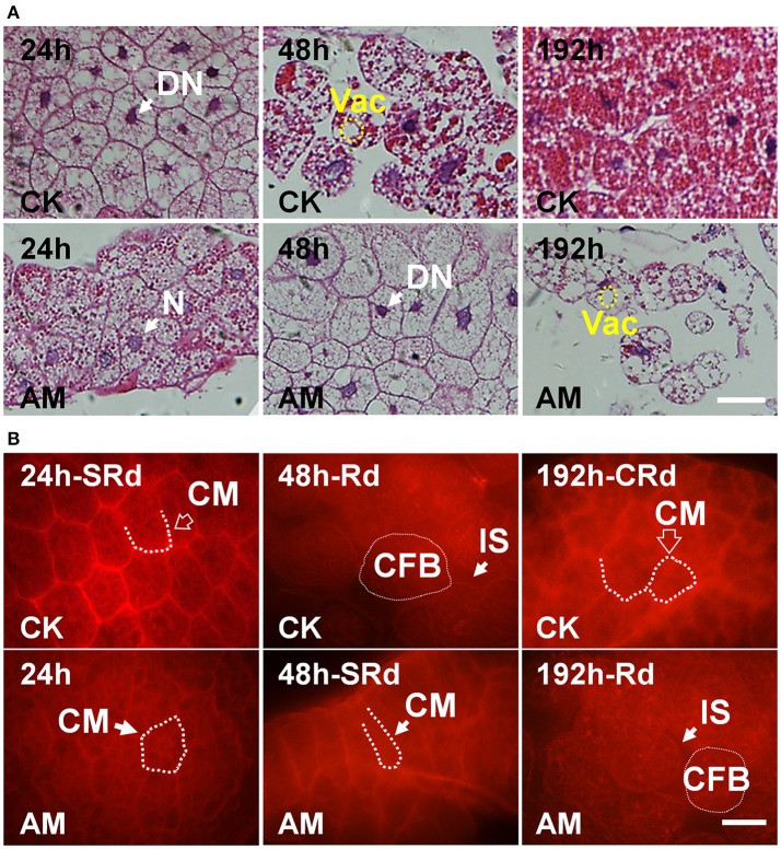
Figure 4.
(A) Hematoxylin-eosin staining and (B) cell membrane staining of the silkworm fat body (FB). Bombyx mori larvae were treated and sampled as described in the Figure 1. CK, the control group; AM, animal model of hyperproteinemia. FBs were analyzed at 24, 48, and 192 h after hyperproteinemia was induced. N, normal nuclei; DN, dense nuclei, CM, cell membrane; Vac, vacuolation; CFB, FB cells; IS, intercellular space; SRd, start of FB remodeling; Rd, remodeling of FB; CRd, complete remodeling of FB. The bar = 5 μm.

