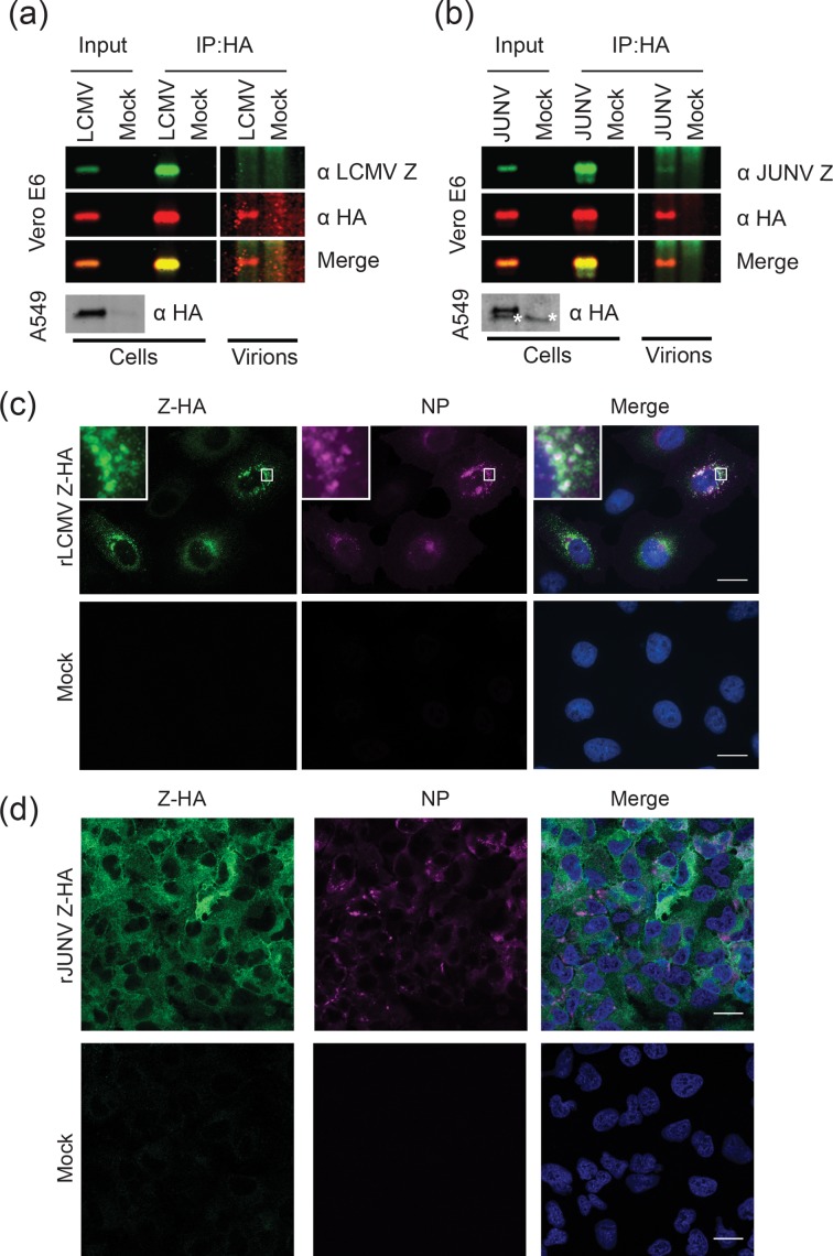
Fig. 2.
Biochemical validation and visualization of arenaviruses encoding an epitope-tagged Z protein. (a, b) Vero E6 or A549 cells were infected with rLCMV Z-HA (a) or rJUNV Z-HA (b) at an m.o.i. of 0.01. Three days later the cells (Vero E6 and A549s) and virus-containing media (Vero E6) were collected and lysed in Triton lysis buffer (Input). HA-tagged Z protein from Vero E6 cells was immunoprecipitated (IP) from cells and virus-containing supernatants using a mouse anti-HA antibody (Covance, MMS-101P) and protein G-coated magnetic beads. Z was detected by Licor two-colour fluorescent Western blotting using antibodies to HA and rabbit immunosera to either LCMV Z (a) [antibody (880), generously provided by M.J. Buchmeier] or JUNV Z (b) (generously provided by Dr Sandra Goñi [46]). A background band detected in A549 cells by the HA antibody is identified by an asterisk. (c, d) Single slices of either mock- or rLCMV Z-HA-infected (c) or rJUNV Z-HA-infected (d) A549 cells at 48 h p.i. that were stained for viral nucleoprotein (NP) (mouse anti-LCMV NP 1–1.3; BEI Resources, mouse anti-JUNV NP NA05-AG12) and HA (Abcam, ab9110) are shown. The perinuclear region of interest in rLCMV Z-HA-infected cells (white box) is enlarged (inset). Scale bars, 20 µm.

