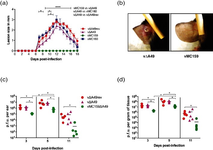
Fig. 3.
The effect of MCV genes on virus virulence using the intradermal (ID) route of inoculation. C57BL/6 mice (n=5 per group) were infected ID with 104 p.f.u. vΔA49, vΔA49rev, vMC159, or vMC160 in the left ear pinna. Lesion size was expressed as the mean for the group +/−SEM. (a) The sizes of the resulting lesions were measured daily for 18 days. The lesion size was measured by using a 0.01 mm digital caliper. The data are expressed as the means of lesion sizes±SEM. Statistical significance was determined by two-way analysis of variance (ANOVA), followed by Tukey’s multiple comparison test. The asterisks indicate the days on which the lesion size caused was statistically significantly between indicated groups (*P<0.05 or ****P<0.0001). (b) Representative images of inoculated ear pinnae at 10 days p.i. vΔA49-infected mice (left panel) or vMC159-infected mice (right panel). (c, d) At the indicated days p.i., ears were collected, homogenized and lysed, and the viral titres of the lysates were determined by plaque assay. Each symbol represents the virus titre from an individual animal, and the mean titre is indicated by a line. The data are expressed as the mean titre of virus (p.f.u.) per gram of tissue (d) and as the total p.f.u. per ear (c). Statistical significance was determined by the Kruskal–Wallis test. The asterisks indicate data points at which the titres of the viruses were statistically significantly different from the others (*P<0.05).

