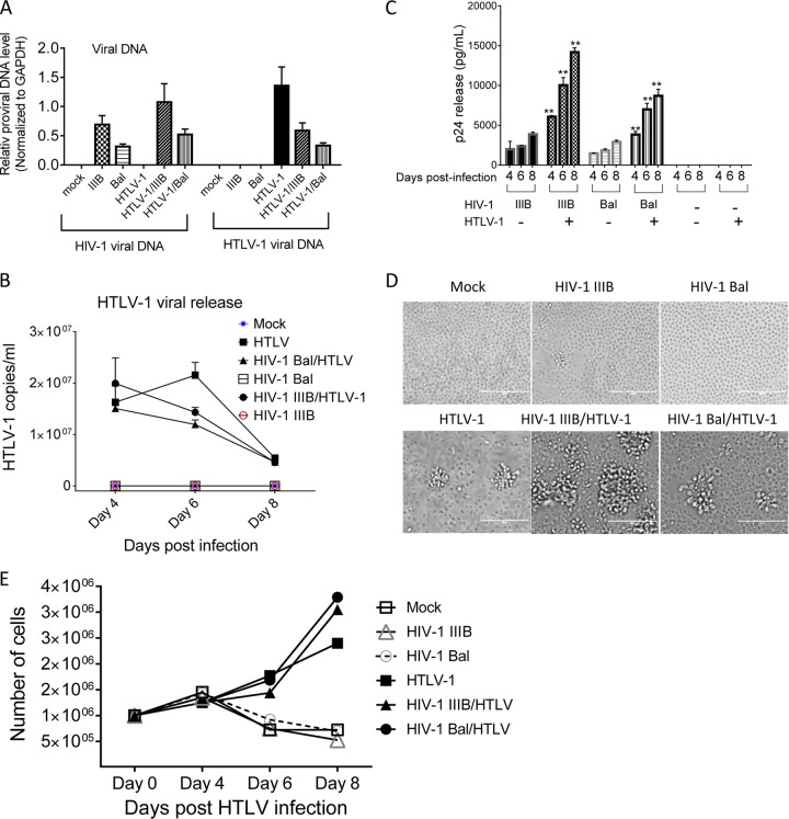
FIG 1 .
Coreplication of HIV-1 and HTLV-1 in primary CD4+ T cells. Primary CD4+ T lymphocytes were infected with HIV-1 IIIB or Bal and/or HTLV-1 as indicated. (A) HIV-1 IIIB or Bal and HTLV-1 DNA in infected cells at day 4 (HIV-1) or 5 (HTLV-1) postexposure was quantified by qPCR. (B) HTLV-1 production in culture supernatant at days 4, 6, and 8 postinfection was determined by qRT-PCR. (C) HIV-1 release at days 4, 6, and 8 postinfection was quantified by p24 ELISA. **, P < 0.001 (in comparison with data obtained with cells infected with HIV-1 alone). (D) Representative images show that exposure to HTLV-1 resulted in morphological changes in primary CD4+ T lymphocytes. Images were taken at day 5 after HTLV-1 exposure. (E) HTLV-1 exposure stimulated primary CD4+ T lymphocyte proliferation. The data shown represent the mean of data from three to six independent experiments.

