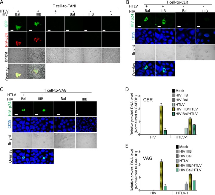
FIG 2 .
Infection of epithelial cells with HIV-1 from HIV-1–HTLV-1-coinfected CD4+ T cells. TANI reporter cells and primary epithelial cells isolated from VAG and CER biopsy tissues were infected by coculture with HIV-1 (Bal or IIIB)- and HTLV-1-coinfected (HTLV+), HIV-1-monoinfected (HTLV−), or mock-infected (HTLV− HIV−) primary CD4+ T cells. The viruses used to infect donor primary CD4+ T cells are indicated in each panel. (A) HIV-1 infection of TANI reporter cells as indicated by GFP expression (green) and HIV-1 Gag expression (red) after immunostaining with anti-HIV-1 Gag antibody. (B, C) HIV-1 infection of primary CER (B) and VAG (C) epithelial cells. At day 5 postinfection, staining was performed with anti-HIV-1 Gag (green) and anti-CK19 (blue) antibodies, followed by confocal immunofluorescence microscopy analysis. The bright and merged views of the same field are shown (bottom). (D, E) DNA levels of both retroviruses in infected primary CER (D) and VAG (E) epithelial cells at day 5 postexposure were determined by qPCR. The data shown represent the mean ± the standard deviation from three independent experiments. Scale bars, 20 µm.

