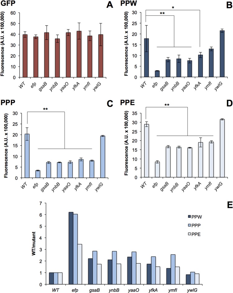
FIG 5 .
A GFP (A), PPW-GFP (B), PPP-GFP (C), or PPE-GFP (D) reporter construct was chromosomally inserted into amyE in each mutant (Rajkovic et al. [22]). After a 1-h induction with 1 mM IPTG, GFP fluorescence was measured. Fluorescence levels were normalized to OD600. Values are means ± standard deviations (SD) (error bars) from three biological replicates. Statistical significance was determined with an analysis of variance (ANOVA) and Tukey posthoc test (*, P < 0.05; **, P < 0.01). (E) Ratio of WT/mutant for each PPX-GFP reporter in each mutant background.

