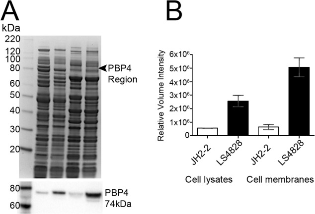
FIG 5 .

Quantitation of PBP4 in the lysate and membrane fractions of JH2-2 and LS4828 shows increased expression in strain LS4828. (A, top) Coomassie-stained 4 to 12% NuPAGE gel and Western blot analysis of E. faecalis samples. Lanes: 1, MagicMark XP Standards; 2, JH2-2 cell lysate; 3, LS4828 cell lysate; 4, JH2-2 membranes; 5, LS4828 membranes. (A, bottom) Western blot analysis of Coomassie-stained gel (above) with polyclonal PBP4 antibody. (B) Analysis of a Western blot assay by densitometry shows 4- and 7-fold increases in PBP4 expression from LS4828 cell lysates and membranes, respectively, and is representative of two independent experiments.
