
FIG 6 .
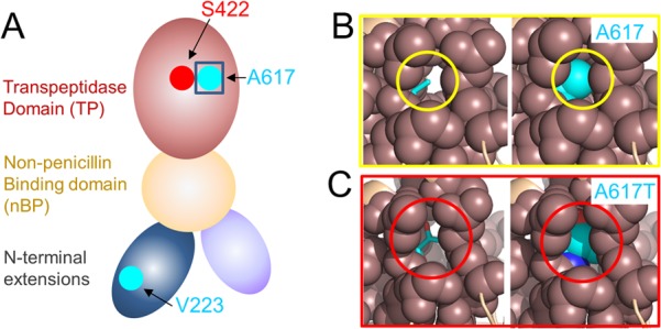
A617 defines the center of an extensive hydrophobic pocket that is adjacent to the PBP4 catalytic pocket. (A) Model of PBP4 based on the domain organization of PBP2a. The locations of the catalytic serine (S424; red) and the two residues mutated in PBP4 from LS4828, V223 and A617 (both cyan), are shown. (B, left) A617 (cyan, sticks) in a model of PBP4 based on the structure of S. aureus PBP2a (PDB code 1MWR; generated with FFAS), with the residues within 5 Å of A617 shown as spheres (coral). (B, right) Same as panel A, except that A617 is shown as spheres. (C, left) PBP4 with the A617T mutation, illustrating the increased space occupied by A617T in this hydrophobic pocket. (C, right) Same as panel B, except that A617T is shown as spheres.