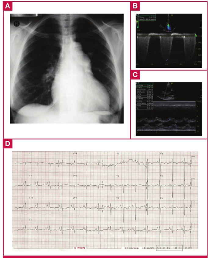
Fig. 3.

ECG of a 38-year-old HIV-positive woman from the PAPUCO cohort. The patient had been on highly active antiretroviral therapy for three years and presented with palpitations and WHO functional class stage III shortness of breath. The chest X-ray (A) shows mild right heart enlargement and borderline raised cardiothoracic ratio. Doppler echocardiographic images (B, C) confirm the diagnosis of severe PH with both severely enlarged right atrium and ventricle with estimated RVSP of 63 mmHg. The ECG (D) shows a normal heart rate and sinus rhythm, right heart enlargement indicated by right-axis deviation of the QRS complex and by a R/S ratio in lead V1 of > 1 with poor R-wave progression. Right ventricular function was altered with a tricuspid annular plane systolic excursion (TAPSE) of 9 mm. Left ventricular ejection fraction was preserved, there was no valvular heart disease and the pericardium was normal.
