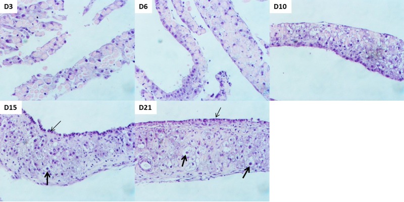
Figure 1.
Organization of cells in organoid culture. Representative photomicrographs of hematoxylin and eosin (H&E)-stained tissue samples obtained over a time course of 21 days. Biliary cells with smaller cuboidal nuclei appear at the surface layer (thin arrows), whereas hepatocytes with bigger round nuclei form the intermediate later (thick arrows). Original magnification: 200×.

