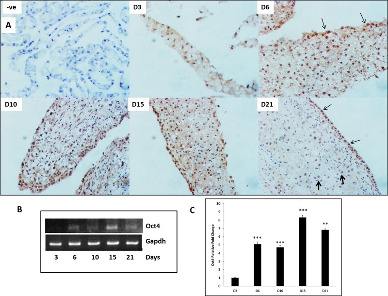
Figure 3.
Oct4 expression during HBT over a time course of 21 days in organoid culture. (A) Representative photomicrographs of Oct4 immunohistochemistry in organoid culture tissue samples. –ve represents “no primary antibody” control. Thin arrows indicate Oct4+ nuclei undergoing HBT. Thick arrows indicate Oct4− nuclei that retain hepatocytic phenotype. Original magnification: 200×. (B) Representative blots of polymerase chain reaction (PCR) product and (C) mRNA levels assessed by quantitative reverse transcription (qRT)PCR) and expressed as fold change relative to GAPDH. Significantly different from day 3 (D3) time point: **p ≤ 0.01, ***p ≤ 0.001.

