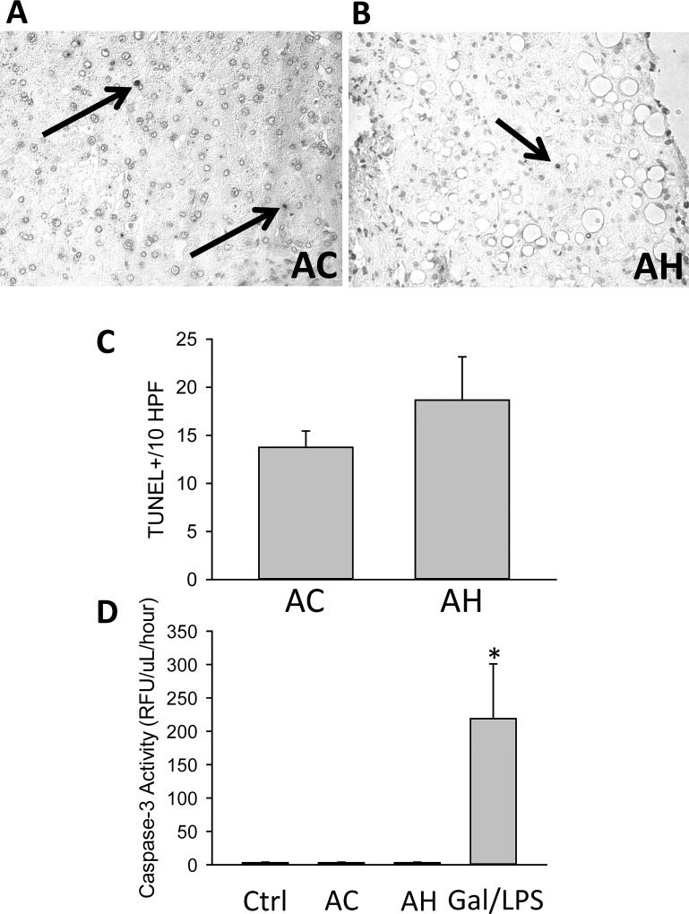
Figure 4.
Cell death markers in patients with AH and AC. TUNEL staining was performed in liver sections from (A) AC and (B) AH patients. Arrows indicate TUNEL+ cells. (C) The numbers of TUNEL+ cells in 10 high-power fields were quantified. (D) In addition, serum caspase 3 activities were measured in HC and in AC and AH patients. Serum from galactosamine/LPS-treated mice was used as a positive control.

