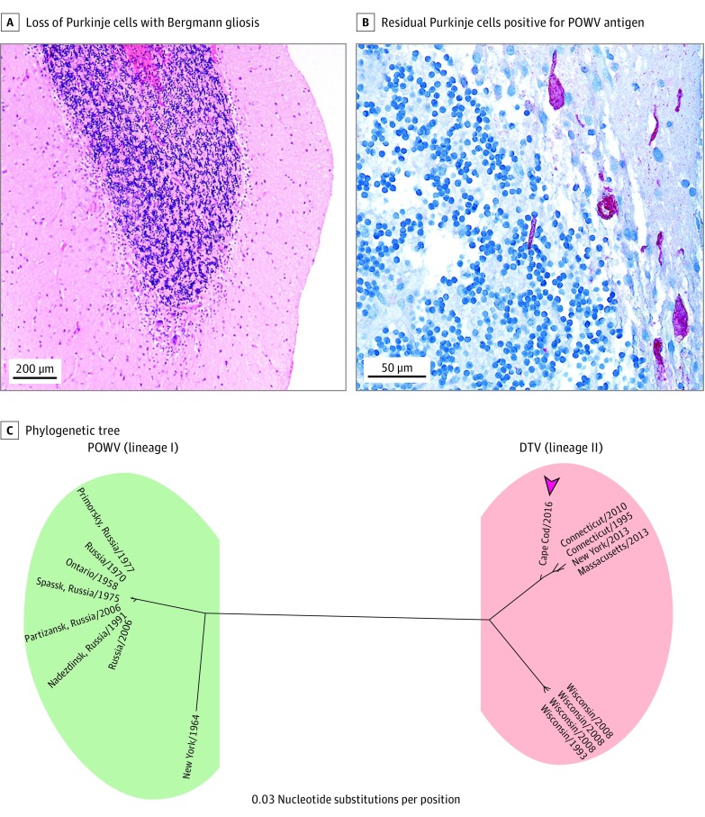
Figure 2. Histological Findings From Cerebellar Biopsy and Phylogenetic Tree.
A and B, Biopsy specimens of cerebellar cortex reveal marked loss of Purkinje cells with Bergmann gliosis (hematoxylin-eosin, original magnification x4) (A) and residual Purkinje cells positive for Powassan virus (POWV) antigen (red) (anti-POWV antibody immunohistochemistry, original magnification x40) (B). C, The 1032-nucleotide segment (nucleotide genome position 9091 to 10 123) of the deer tick virus (DTV) (lineage II) nonstructural protein 5 gene amplified from the patient’s cerebrospinal fluid (Cape Cod/2016 [arrowhead]) was compared against 18 POWV (lineage I) and DTV whole genomes using the neighbor-joining method, demonstrating that this patient’s strain is temporally and geographically most proximate to the DTVs circulating in the northeastern United States over the past 20 years. The phylogenetic tree was produced using a software program (Geneious, version 10.1.3; Biomatters Limited). GenBank accession numbers for the viruses included in the phylogenetic tree are listed in the eTable in the Supplement.

