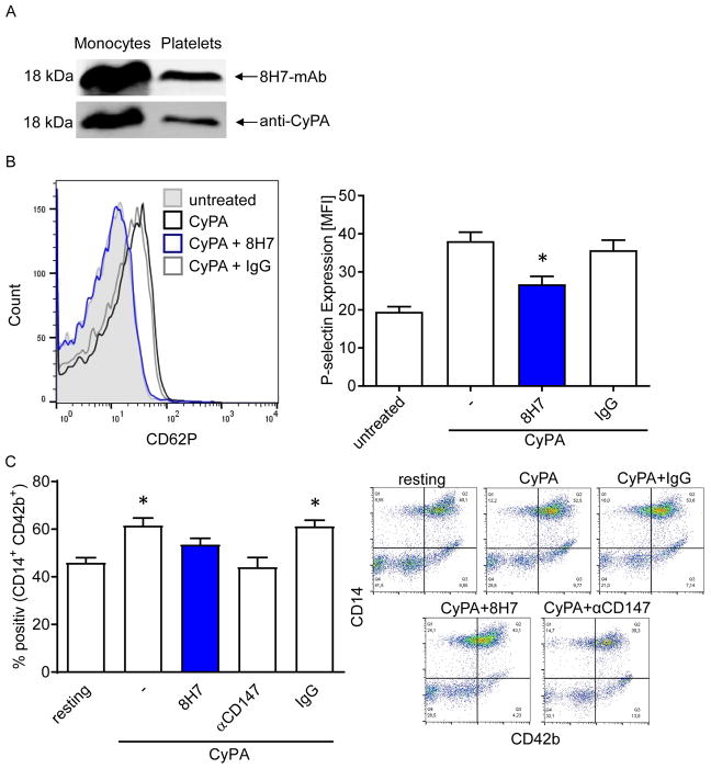
Figure 4. Extracellular CyPA enhances platelet-activation in vitro.
(A) 8H7-mAb detects CyPA in monocyte as well in platelet lysate (upper line). Afterwards the same membrane was incubated with an anti-CyPA antibody (lower line) to prove the specificity of 8H7-mAb. (B) 8H7-mAb significantly reduces the CyPA-dependent platelet activation. Representative overlays of the p-selectin expression and bar graphs show mean±SEM of P-selectin expression on platelets. * means p ≤ 0.05 vs. CyPA+IgG. (C) Monocyte-platelet aggregate (MPA) formation was analyzed by using double staining with CD42b (as platelets marker) and CD14 (as monocyte marker) as described in material and methods. The platelets were treated as indicated and incubated with monocytes for the MPA formation. Figure shows representative flow cytometry analysis. (n ≤ 6) * means p ≤ 0.05 vs. resting. Right panel shows representative quadrat statistic for the CD14+CD42+ double positive cells.

