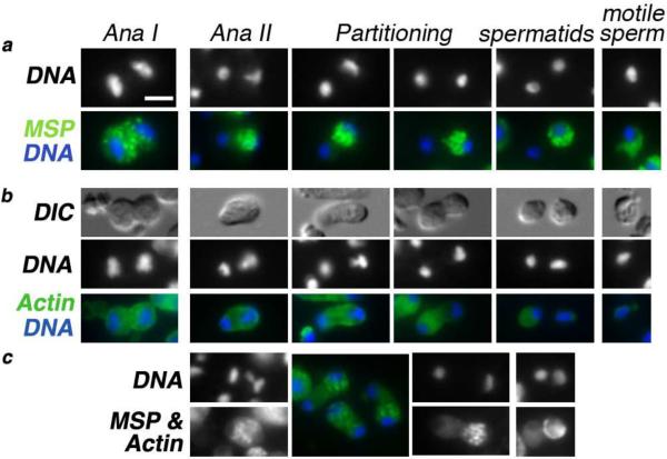
Figure 3. Differential partitioning of cellular components following anaphase II.
(a) Localization and partitioning of the major sperm protein (MSP) relative to the stage of spermatogenesis. Immunostaining of MSP (green), DAPI-stained DNA (white or blue). During the meiotic division and the partitioning process, MSP exists in a punctuate fibrous body state. In motile spermatozoa, it stains uniformly throughout the pseudopod but is absent in the cell body. (b) Localization and partitioning of actin (green) to the sperm with the smaller chromatin mass. DNA (white or blue). (c) In co-stained cells, using a single anti-mouse secondary antibody, the punctuate MSP fibrous bodies partition to the sperm with the X and the large chromatin mass while the smooth and less intense actin staining pattern is associated with the other spermatid. Arrow points to a rare instance of symmetric MSP partitioning. All images are at the same scale; scale bar = 3 μm.

