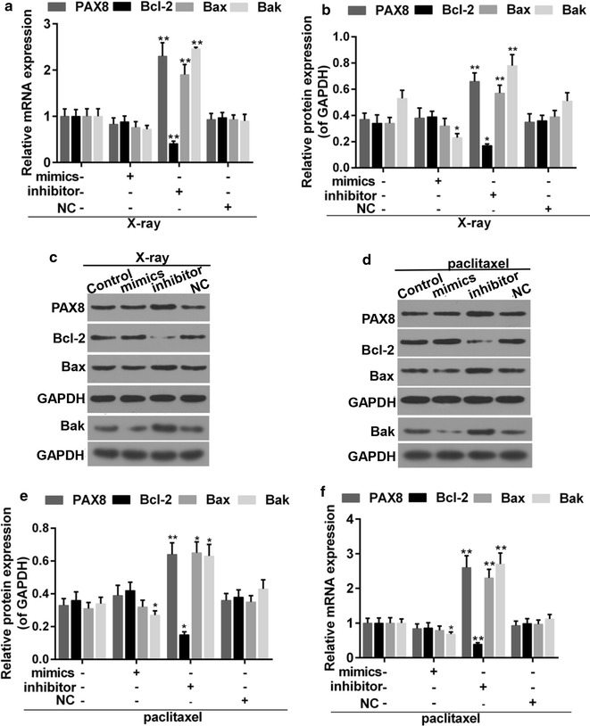
Fig. 8.

a Quantitative analysis was carried out for expressions of PAX8, Bax, Bcl-2 and Bak in PTC cells that were treated with X-ray and miR-144-3p. b, c Western blot was performed for expression of PAX8, Bax Bcl-2 and Bak. d, e Western blot was used for expressions of PAX8, Bax Bcl-2 and Bak in PTC cells that were treated with paclitaxel and miR-144-3p. f Quantitative analysis was performed for expression of PAX8, Bax Bcl-2 and Bak. *P < 0.05, **P < 0.01 vs. control
