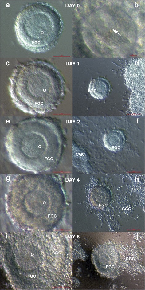
Fig. 4.

In vitro maturation of the immature follicle: the day of retrieval (a) of an oocyte that expressed a germinal vesicle (arrow) (b); on day 1, a slightly thicker layer of granulosa cells around the oocyte (c) in the co-culture with granulosa cells that approached the follicle (d); on day 2, a significantly thicker zona pellucida (e) in the co-culture with granulosa cells that spread over the follicle (f); on day 4, a thicker layer of granulosa cells around the oocyte (g) in the co-culture with granulosa cells, which started to incorporate into the layer of the granulosa cells around the oocyte (h); and on day 8, a massive layer of granulosa cells around the oocyte (i) in the co-culture with granulosa cells, which was massively incorporated into the layer of granulosa cells around the oocyte (j). (Inverted microscope, magnification 200× for b, 100× for a, c, e, g, i, and 40× for d, f, h, j). Legend: O-oocyte, FGC-granulosa cells around the oocyte in the follicle and CGC-co-cultured granulosa cells retrieved by denudation of oocytes in the patient. Red Bar: 10 μm for b, 50 μm for a, c, e, g, i, and 100 μm for d, f, h, j
