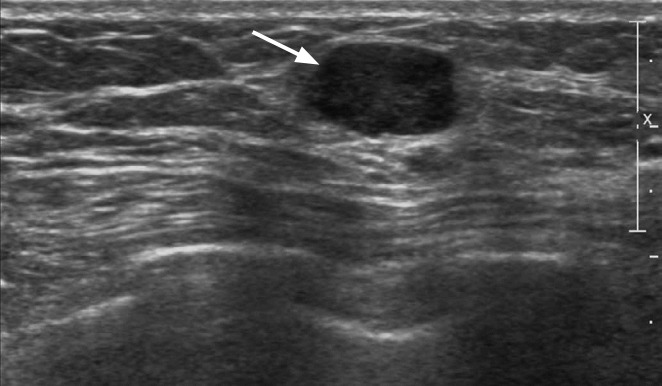
Fig. 2. A 68-year-old woman with a palpable mass in the left breast.

Ultrasonography (US) shows a circumscribed, oval-shaped, hypoechoic mass (arrow) with posterior acoustic enhancement in the left breast. A US-guided biopsy confirmed triple-negative breast cancer.
