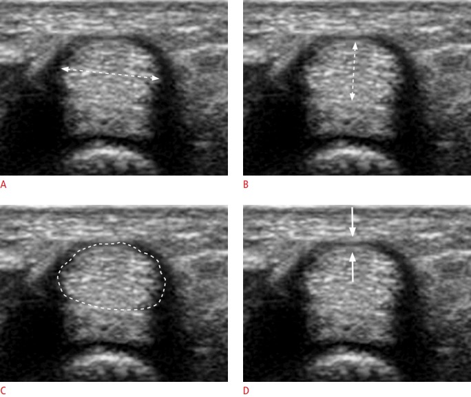
Fig. 2. Axial view for the measurements.

A-D. An axial view on ultrasonography is used in this study to measure four parameters: the transverse diameter of the tendon (A, dotted line), the thickness of the tendon (B, dotted line), the cross-sectional area of the tendon (C, dotted line), and the thickness of the pulley (D, arrows). The tendon shows a hyperechoic fibrillar pattern, while hypoechoic fluid distension is maximal.
