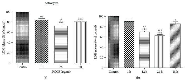
Figure 7.
Effects of PCGE on H2O2-induced LDH release from human primary astrocytes. (a) Cells were preincubated for 24 h with the different concentrations of PCGE (15, 25, and 50 μg/ml) and then exposed to 0.5 mM of H2O2 for 4 h in FBS-free medium. ∗∗,∗∗∗Significant differences at the P < 0.01 and P < 0.001 levels, respectively, versus the control group; #P < 0.05 versus the 15 μg/ml group. (b) LDH release from astrocyte cells that were pretreated with 25 μg/ml PCGE for different durations (up to 48 h) before exposure to 0.5 mM H2O2. ∗,∗∗∗Significant differences at the P < 0.05 and P < 0.001 levels, respectively, versus the control group; ##P < 0.01 and ###P < 0.001 versus the 1 h group. The results were quantified by using an LDH activity kit assay and expressed as percentages of LDH released in PCGE-treated or vehicle-treated cells (control) exposed to H2O2. Values are means ± SE; n = 5 independent experiments. ANOVA followed by the Tukey post hoc test.

