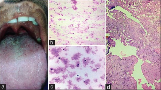
Figure 1.

(a) Swelling in the posterior part of the tongue. (b and c) Smears showed scattered mucinophages and lymphocytes in a mucinous background. (b: Giemsa stain x100; c: Pap stain x400). (d) Histopathology revealed both hypocellular and hypercellular areas with cystic areas. (H and E stain ×100)
