
Extended Data Figure 4. Passive transfer of ZIKV NS1 antibodies into infected AG6 mice prevented ZIKV acquisition by A. aegypti.
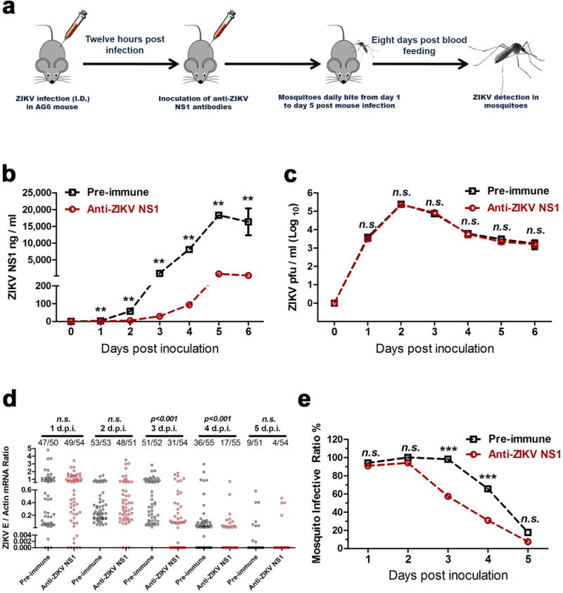
a, Schematic representation of the study design. AG6 mice were intradermally infected with 1×10⁴ pfu of the ZIKV GZ01 strain. Subsequently, 100 μl of a murine ZIKV NS1 antibody was intraperitoneally inoculated into the mice 12 hr post-infection. The same amount of pre-immune serum was inoculated as a mock control. After 12 hr of antibody dissemination, the infected mice were subjected to daily biting by female A. aegypti from day 1 to day 5 post-mouse infection. The mouse blood-fed mosquitoes were reared for an additional 8 days for ZIKV detection.
b, ZIKV NS1 measurement by ELISA (n=6 mice per group pooled from 3 independent biological replicates). Mouse sera were collected to quantify the amounts of ZIKV NS1 protein from days 0 and 6 post-mouse infection.
c, Detection of the ZIKV load in the blood of the infected mice (n=8 mice per group pooled from 4 independent biological replicates). The presence of infectious ZIKV particles in blood plasma was determined by a plaque assay from days 0 to 6 post-mouse infection.
d, e, Immuno-blockade of ZIKV NS1 in infected AG6 mice reduced the infection of fed A. aegypti (n=6 mice per group pooled from 3 independent biological replicates). The number of infected mosquitoes relative to the total number of mosquitoes is shown at the top of each column. Each dot represents a mosquito (d). The data are represented as the percentage of mosquito infection (e).
Data are mean ± s.e.m. (b, c). p values were determined by two-tailed Mann-Whitney test (b-d) or two-sided Fisher’s exact test (e). **p< 0.01, ***p< 0.001, n.s., not significant.