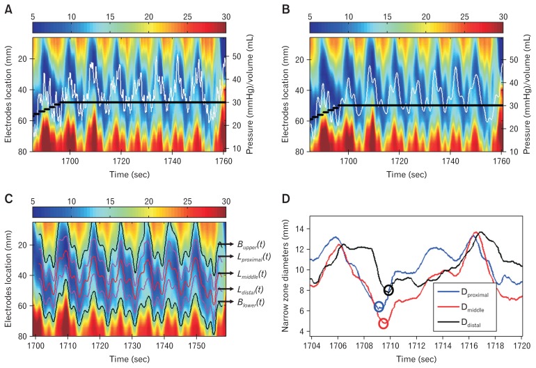
Figure 2.
Example of pressure and diameter change before (A) and after (B) the low-pass filter. By using the low-pass filter, the signals with frequency < 30 cycle/min was passed and unchanged in (B), while the fluctuations due to heart action in (A) was successfully removed after the filter in (B). The white line is the pressure change during the distension and the heavy black line is the volume change in the functional luminal imaging probe bag. (C) The segmented lower esophageal sphincter (LES) narrow zone from the spatial-temporal diameter map shown in (B), where the upper edge (Bupper(t)), lower edge (Blower(t)), proximal (Lproximal(t)), middle (Lmiddle(t)), and distal (Ldistal(t)) locations of the narrow zone were segmented during the distension. (D) Bag diameter during a cycle at the proximal, middle, and distal locations of the LES narrow zone. In the diameter cycle, the minimum diameter at the proximal narrow zone (blue circle) was reached at first and followed by the middle (red circle) and distal (black circle) of the narrow zone.

