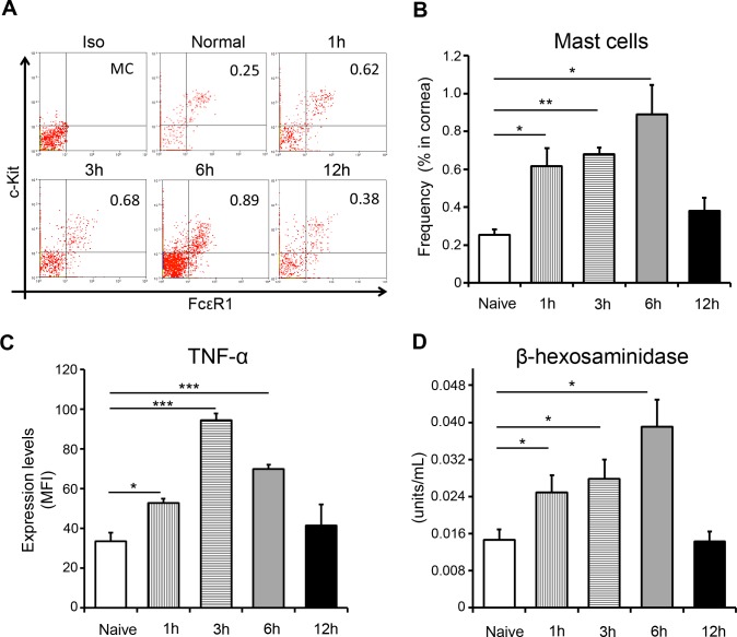
Figure 2.
Corneal injury results in activation of mast cells at the ocular surface. (A) Representative flow cytometric dot plots and (B) cumulative bar chart showing the frequencies of ckit+FcεR1+ mast cells in the cornea at different time points after injury, relative to naïve mice. (C) Bar chart depicting the expression (MFI) of TNF-α by mast cells at different time points after injury, relative to naïve mice. (D) Corneal tissue (with limbus) were lysed, and β-hexosaminidase levels were estimated at different time points after injury, relative to naïve mice (as described in Materials and Methods). Representative data from three independent experiments are shown, and each experiment consisted of four to six animals. Data are represented as mean ± SD. *P < 0.05; **P < 0.01; ***P < 0.001.

