Figure 5.
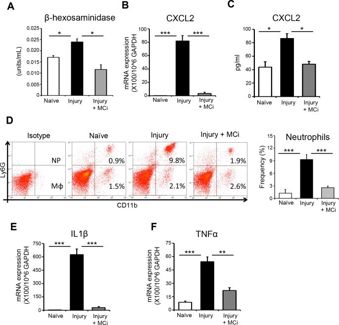
In vivo inhibition of mast cells prevent early recruitment of neutrophil and inflammation at ocular surface after corneal injury. Mast cell inhibitor (MCi) was topically administered to the ocular surface at −6, −3, 0, and 3 hours after corneal injury. Corneas with limbus were harvested at 6 hours after injury. PBS-treated mice with corneal injury and naïve mice served as controls. (A) Bar chart showing β-hexosaminidase levels in lysed corneas in MCi-treated mice, PBS-treated mice, and naïve controls. (B) Bar chart showing CXCL2 expression by mast cells (normalized to GAPDH) in the indicated groups, as quantified by real-time PCR. (C) Bar chart depicting protein levels of CXCL2 in the corneal lysate of indicated groups, as quantified by ELISA. (D) Single cell suspensions were prepared, and flow cytometry was performed. Representative flow cytometry plots (left) and bar chart (right) showing infiltration of CD11b+Ly6G+ neutrophils and CD11b+Ly6G− macrophages to the ocular surface of indicated groups. (E) Bar chart showing expression of IL-1β mRNA in the conjunctivae harvested from indicated groups, as quantified by real-time PCR. (F) Bar chart showing expression of TNF-α mRNA in the conjunctivae harvested from indicated groups, as quantified by real-time PCR. Results are representative of three independent experiments. Each group consisted of five to six animals in each experiment. The values shown represent mean ± SD. *P < 0.05; **P < 0.01; ***P < 0.001.

