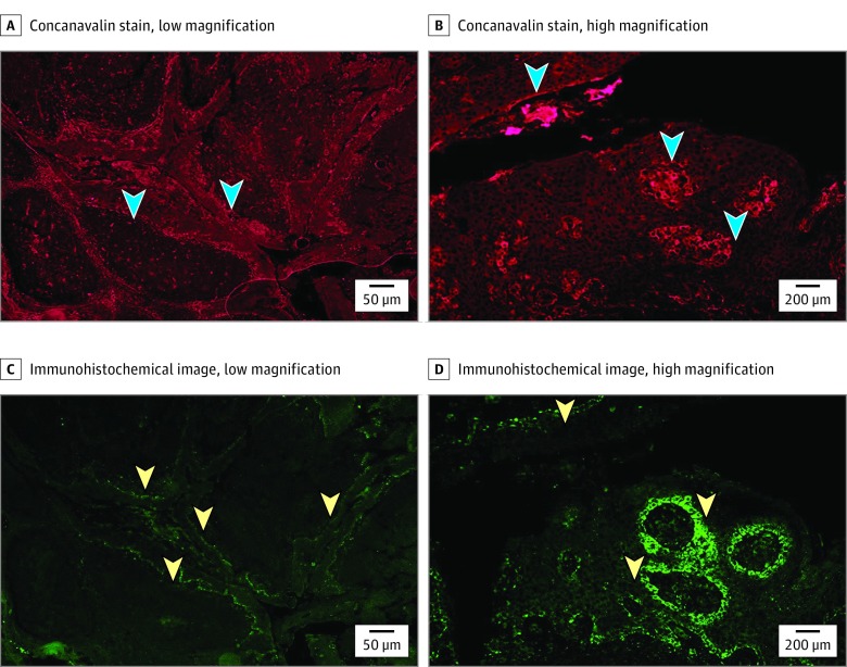
Figure 1. Histopathologic Images.
A, Tonsil tissue stained with concanavalin A (arrowheads indicate glycocalyx matrix, original magnification ×4). B, Tonsil tissue stained with concanavalin A (arrowheads indicate glycocalyx matrix, original magnification ×20). C, Tonsil tissue immunohistochemistry with human papillomavirus (HPV) L1 Ab (arrowheads indicate HPV L1 Ab, original magnification ×4). D, Tonsil tissue immunohistochemistry with HPV L1 Ab (arrowheads indicate HPV L1 Ab, original magnification ×20).

