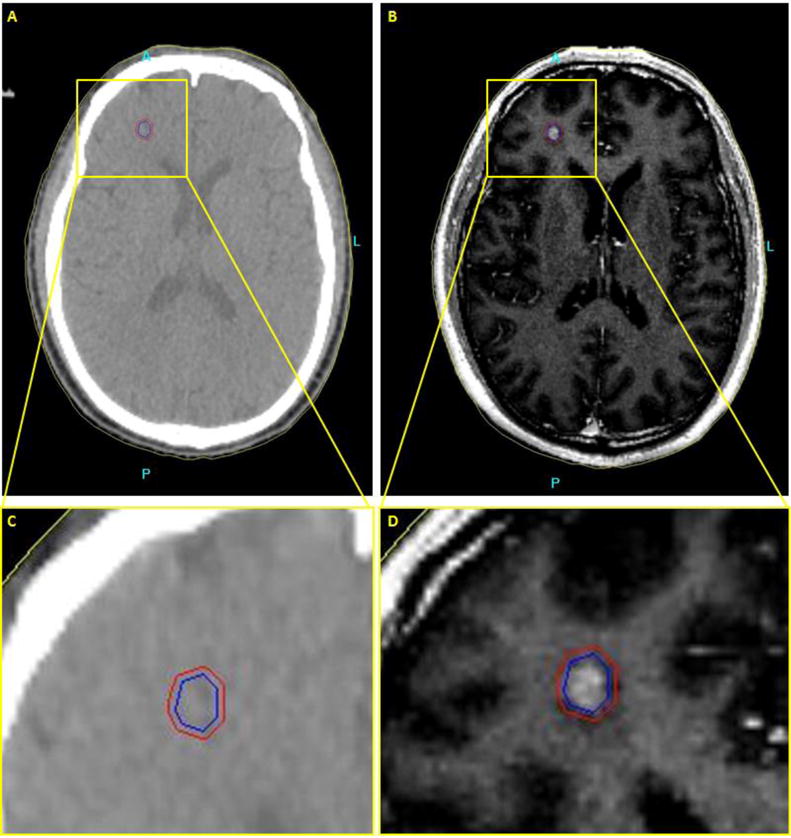
Figure 1.

Axial brain images of patient with a metastatic tumor in the brain. (a,c) CT image. No contrast between the tumor and the surrounding normal tissue. (b,d) T2-weighted Fluid-attenuated inversion recovery image (FLAIR) MR image. Higher soft tissue contrast of the MR image leads to more accurate delineation of the tumor.
