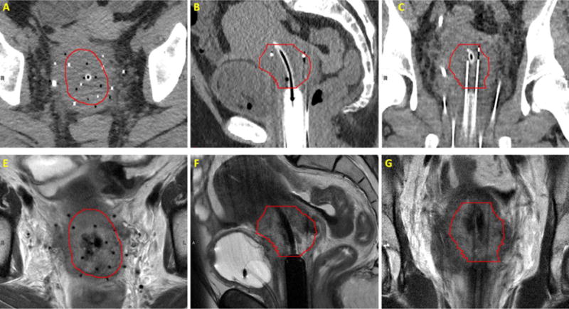
Figure 3.

Comparison of CT and T2-weighted images of a patient with plastic needles and a plastic cylinder and tandem in place; Transverse view of CT (top row) and T2-weighted (bottom row) of a patient’s pelvis with axial view showed in left panel and sagittal and coronal views showed in middle and right panels, respectively. High-risk CTV volume shown in red.
