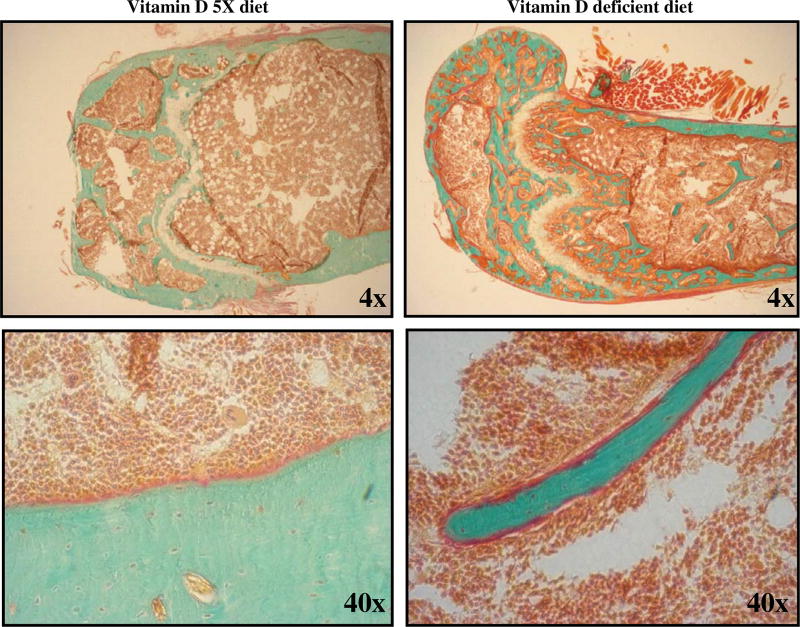
Fig. 2.
Effects of vitamin D deficiency on bone histology. Goldner’s trichrome stain for 7 µm thick sections of non-demineralized bone embedded in methyl methacrylate: mineralized bone stains green. Unmineralized matrix or osteoid along the mineralized bone periphery stains red. Growth plate cartilage stains light-gray and chondrocytes light-red. Bone marrow cells stain red and marrow adipocytes do not stain. Femur tissue sample from mouse fed a vitamin D-enriched diet (Vitamin D 5X diet, left column) shows few, very thin osteoid layers or seams. Femur tissue sample from mouse fed a vitamin D-deficient diet (Vitamin D deficient diet, right column) shows a thickened osteoid layer, an abnormality indicating Vitamin D deficiency.

