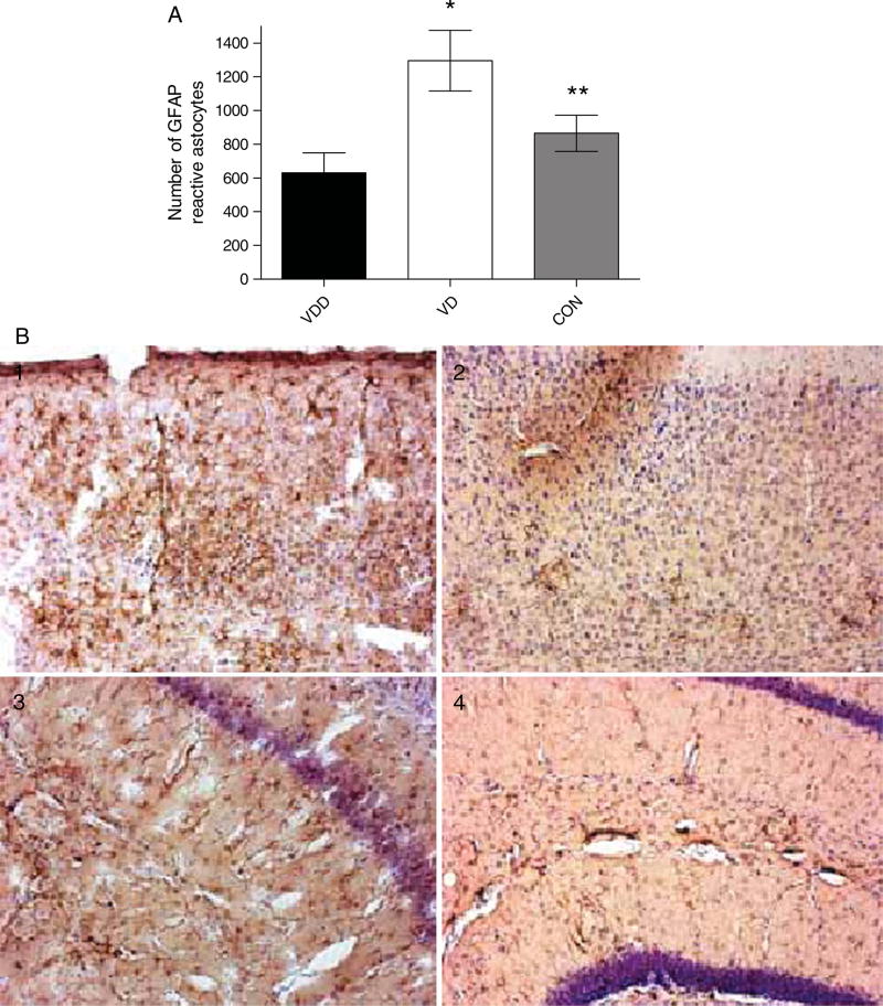
Fig. 8.
GFAP immunoreactivity in the brains of AβPP mice treated with vitamin D. Brain sections of AβPP mice were immunostained with anti-mouse GFAP antibody to detect the distribution of activated astrocytes. A) AβPP mice fed a vitamin D-enriched diet (VD, white bars) versus AβPP mice fed a vitamin D-deficient diet (VDD, black bars). B) Examples of sections showing activated GFAP-positive astrocytes. 1 and 2, cortex; 3 and 4 hippocampus from AβPP mice on vitamin D-enriched diet (1 and 3) or vitamin D-deficient diet (2 and 4). Data are presented as mean ± S.E. *p < 0.001; **p < 0.01 (n = 10 per group).

