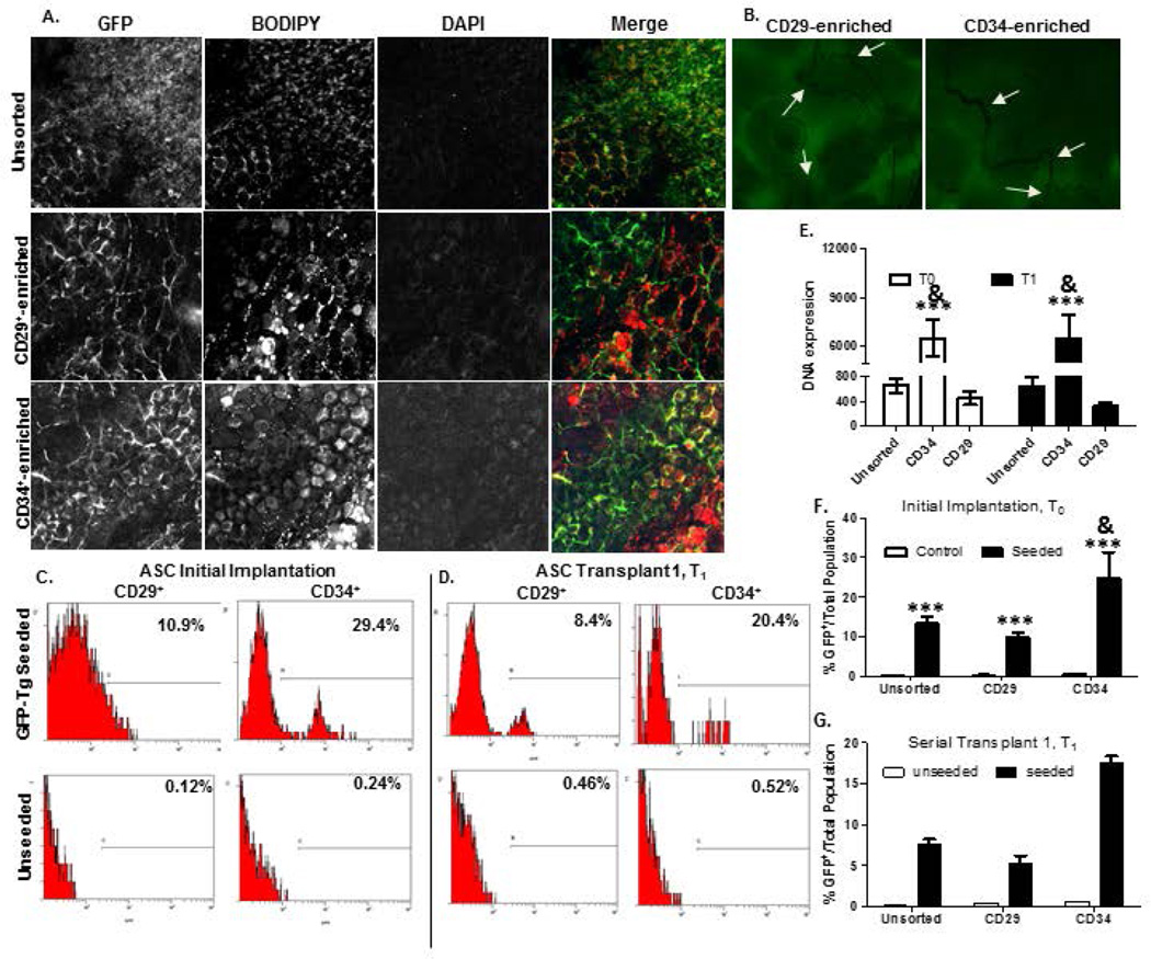
Figure 6. Detection of CD146− CD29+ GFP-Tg ASC and CD146− CD34+ GFP-Tg ASC from tissue engineered adipose using HFIP 6-week silk scaffold implants in mice over two serial transplantations.
Photomicrographs of GFP-Tg CD29-enriched and CD34-enriched (cohorts (a)-(c) cell implantation following week 6 in vivo demonstrate persistence, proliferation, and ability to form GFP-Tg adipose depots, similar to unsorted GFP-Tg SVF cohorts. 6a). Confocal microscopy images of GFP, BODIPY, DAPI, and merged images from 6-week GFP-Tg ASC serial (T1) transplants. 6b) Confocal images of CD29-enriched and CD34-enriched 6-week T1 groups reflected observation of micro vessel formation within CD34-enriched groups than in CD29-enriched groups; n=5 per group per replicate. Flow cytometric analyses of GFP expression in removed adipose scaffolds from cohorts (a)-(c) following 6c) T0, and 6d) T1 transplants. 6e). GFP DNA expression in samples from removed 6-week T0 and T1 transplants. GFP expression reported as fold expression and normalized to GAPDH expression. Quantification of %GFP positivity following 6-week transplantation in 6f) T0 and 6g) T1 GFP-Tg ASC cell implants. Data reported as mean + SD. Comparing individual grouping to unsorted cell seeded groups: *p < 0.05, ** p < 0.01; ***p < 0.001; comparing to CD34-enriched group: & p < 0.05

